The Lung
- Bronchioles – lack of cartilage and submucosal glands in walls
- Terminal bronchioles – less than 2mm in diameter
- Acinus – lung distal to terminal bronchiole. Spherical around 7mm in diameter. Respiratory bronchiole with some alveoli –> alveolar duct –> alveolus.
- Pulmonary lobule – cluster of 3-5 terminal bronchioles with acini
- Whole resp epithelium except vocal cords are covered by pseudostratified tall columnar ciliated epithelial cells.
- Mucus secreting goblet cells in cartilagenous airways.
- Neuroendocrine cells – seretonin, calcitonin, gastrin releasign peptide.
- Alveolar walls – capillary endothelium–> basement membrane–> alveolar epithelium
- Type 1 – 95%
- Type 2 – make surfactant, repair alveolar epithelium
Atelectasis.
- Incomplete expansion or collapse of previously expanded lung.
- Resorption(obstruction) – mediastinum shift ipsilaterally
- Compression – pneumothorax, haemothorax, tumour, pus, pleural effusion – mediastinum shift contralaterally
- Contraction – from scarring/fibrosis
- Reduces oxygenation and increases risk of infection.
Pulmonary oedema
- Congestive(cardiogenic)
- Haemosidirin laden macrophages, fibrosis –> firm brown–> impair resp function and increase rate of infection
- Increased capillary permeability(microvascular injury to alveolar septa vessels)
- Leakage of protein and fluids into interstitum then alveoli
- Usually localised – pneumonia
- If generalised – life threatening ARDS
ARDS (Acute Respiratory Distress Syndrom)
- Clinical syndrome caused by diffuse alveolar capillary injury–> vasc permeability–> alveolar flooding–> decreased diffusion–> surfactant abnormalities, exudate –> scarring
- Chemotaxis – IL8, TNFa, IL-1 – neutrophil predominant
- Rapid onset severe resp failure
- cyanosis
- Hypoxaemia refractory to O2 therapy
- Possible multi organ failure
- CXR – diffuse alv infiltrate
- 60% mortality
Pulmonary embolism
- Pulmonary emboli originate from deep venous thromboses and are the most common form of thromboembolic disease.
- Fragmented thrombi from DVTs are carried through progressively larger veins and the right side of the heart before slamming into the pulmonary arterial vasculature.
- Depending on the size of the embolus, it can
- occlude the main pulmonary artery
- straddle the pulmonary artery bifurcation (saddle embolus), or
- pass out into the smaller, branching arteries
- Most pulmonary emboli (60% to 80%) are clinically silent because they are small. With time they become organized and are incorporated into the vascular wall; in some cases organization of the thromboembolus leaves behind a delicate, bridging fibrous web.
- Sudden death, right heart failure (cor pulmonale), or car diovascular collapse occurs when emboli obstruct 60% or more of the pulmonary circulation.
- Embolic obstruction of mediumsized arteries with sub sequent vascular rupture can result in pulmonary hemorrhage but usually does not cause pulmonary infarction. This is because the lung is supplied by both the pulmo nary arteries and the bronchial arteries, and the intact bronchial circulation is usually sufficient to perfuse the affected area. Understandably, if the bronchial arterial flow is compromised (e.g., by leftsided cardiac failure), infarction may occur.
- Embolic obstruction of small endarteriolar pulmonary branches often does produce hemorrhage or infarction.
- Multiple emboli over time may cause pulmonary hypertension and right ventricular failure.
Chronic obstructive pulmonary disease.
- Emphysema(pink puffer) and chronic Bronchitis(blue bloater) – obstructive diseases
- Loss of elastic recoil of lung therefore reduced Expiratory flow.
- Of patients 10% are non smokers. Only a minority of smokers develop it.
- Emphysema – abnormal permanent enlargement of airspaces distal to the terminal bronchiole, associated wall destruction without fibrosis.
- Centriacinar (95%)- apical, in heavy smokers.
- Pan acinar – basal, in a1antitrypsin deficiency
- Paraseptal – spont pneumothorax
- Irregular – fibrosis
- Pathogenesis –
- protease-antiprotease theory – smoking causes inhibition of antiproteases therefore results in imbalance between proteases and elastases and their inhibitors leading to increased wall destruction. They also cause chemotaxis and cytokines cause further O2 free radical damage and increase macrophage elastase and metalloproteinases. So there is also an oxidant/antioxidant imbalance.
- Clinical manifestations start when 1/3 of parenchyma damaged. DYSPNOEA, weight loss, pursed lips, barrel chest.
- Needs spirometry to diagnose and quantify the disease
- Chronic bronchitis – recurrent infections, prominent cough and sputum(goblet cell metaplasia). Hypercapnoea, hypoxaemia–> cor pulmonale–> CCF
- Secondary infections due to smoking intefering with cilliary function, leukocyte function and causing direct epithelial damage.
Asthma.
- Airway hypersensitivity(bronchospasm) and inflammation –> decreased airway diameter –> obstruction to flow.
- Chronic inflammatory disorder of airways causing recurrent wheezing, dyspnoea, chest tightness and cough.
- Eosinophils, IL4, IgE –> TH2 predominence.
- Immediate phase – Ag-Ab cross linking –> IgE release and Mast cells degranulate–> histamine bronchoconstricts and opens tight junctions between epithelial cells–> antigen enters and activates further mast cells, eosinophils, –> further mediators –> bronchospasm, inc vasc perm and inc mucus production, and chemotaxis
- Late phase–> due to the chemotaxis there is a fresh round of mediator release and inflammation after the initial trigger has been removed.
- Histamine, prostaglandin D2, PAF, leukotrienes
- Morphology – mucus plugs, whorls of epithelium(Curschmann spirals), eosinophils, basement membrane thickening, oedema, inc size of submucosal glands, bronchial wall muscle hypertrophy.
- Lung function tests
- FEV1 – reduces in obstructive lung problems and increases in restrictive lung problems
- FVC – reduces in restrictive lung issues where lungs cannot inflate enough.
- Ratio FEV1:FVC in COPD is <70%
Bronchiectasis.
- Permanent Dilation of bronchi and bronchioles caused by destruction of muscle and elastic tissue resulting from or associated with chronic necrotising infections.
- CF, post infectious, bronchial obstruction
- Clinical: severe persistent cough, haemoptysis
- Cx – obstruction, dyspnoea, cyanosis, cor pulmonale, metastatic brain abscess, amyloidosis
Pneumonia.
- Infection of alveoli
- Community acquired – strep pneumonia, haemphilus influenzae, moraxella catarrhalis, staph aureus, legionella, Klebsiella
- Atypical – Chlamydia pneumoniae, mycoplasma pneumoniae, coxiella brunetti, viral
- Aspiration
- Chronic – Norcadia
- Immunocompromised – CMV, pneumocystis carinii, MAC
- Cx – fever, cough, myalgia, lethargy, SOB, respiratory failure, abscess, empyema, organisation, bacteraemic dissemination.
- Broncho pneumonia vs Lobar pneumonia.
- Lobar has 4 stages
- Congestion
- Red hepatization
- Gray hepatization
- Resolution
- Influenza virus – single stranded RNA bound by nucleoprotein(determines if type a b or c). Lipid bilayer envelope with haemaglutanin and neuraminidase(determine subtype) – cleared by cytotoxic T cells and intracellular anti influenza protein in macrophages.
- Epidemics – due to entigenic drift – mutations of haemaglutinin or neuraminidase
- Pandemics – antigeic Shift – recombination of RNA segments with animal viruses.
Lung Abscesses.
- Local suppurative process in lung characterised by necrosis of lung tissue
- Oropharyngeal surg, sinobronchial infections, dental sepsis and bronchiectasis, aspiration, antecedent bacterial infection, septic emboli, neoplasia can lead to it.
- Streptococci, Staph aureus, GN bacteria, mixed infections.
- Pulmonary TB – Apical
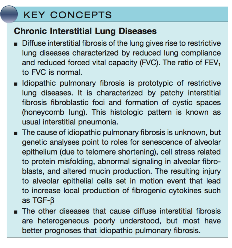
Chronic Diffuse Interstitial (Restrictive) Lung Diseases
Robbins & Cotran Pathologic Basis of Disease 9E (2014)
- Chronic interstitial pulmonary diseases are a hetero- geneous group of disorders characterized predominantly by inflammation and fibrosis of the pulmonary interstitium.
- Many of the entities are of unknown cause and pathogenesis, and some have an intra-alveolar as well as an interstitial component. There is frequent overlap in histologic features among the different conditions. These disorders account for about 15% of noninfectious diseases seen by pulmonary physicians
- Clinical features
- In general, the clinical and pulmonary functional changes are those of restrictive lung disease. Patients have dyspnea, tachypnea, end-inspiratory crackles, and even- tual cyanosis, without wheezing or other evidence of airway obstruction.
- The classic functional abnormalities are reductions in diffusion capacity, lung volume, and lung compliance.
- Chest radiographs show bilateral lesions that take the form of small nodules, irregular lines, or ground- glass shadows, all corresponding to areas of interstitial fibrosis.
- Eventually, secondary pulmonary hypertension and right-sided heart failure associated with cor pulmonale may result.
- Although the entities can often be distinguished in the early stages, the advanced forms are hard to differ- entiate because all result in scarring and gross destruction of the lung, often referred to as end-stage lung or honeycomb lung.
- Diffuse restrictive diseases are categorized based on histology and clinical features
Lung tumours.
- 90-95% carcinomas
- Squamous cell – 25-40%
- Smokers, paraneoplastic sometimes cause hypercalcaemia
- Adenocarcinoma – 25-40%,
- women and non smokers
- Small cell – 20-25%
- Smokers, all are high grade, highly malignant, major bronchi and periphery, metastasise widely.
- Untreated survival time 6-17wks, sensitive to chemo and radiation’
- paraneoplastic sometimes release ADH or ACTH
- Large cell – 10-15%
- Squamous cell – 25-40%
- 5% Bronchial carcinoids
- Paraneoplastic syndromes – symptoms occuring in pt with neoplasm that are not explained by the tumour or its mets and the hormones are not released by the tumour
- ADH, ACTH, PTH, Calcitonin, Gonadotrophins, Seretonin, bradykinin,
Pleural conditions.
- Asbestos alone increases lung cancer rate by 5, with smoking it increases to 55 fold. Asbestos increases mesothelioma to 1000 times but not increased by smoking.
- Pleural effusion – inflammatory or noninflammatory
- Pneumothorax
- Tumours – mesothelioma
Viva questions:
- Classify the causes of pulmonary oedema
- What is ARDS and what conditions can cause it?
- Tell me about the pathogenesis and types of emphysema.
- Tell me about the pathogenesis of chronic bronchitis.
- Tell me about COPD
- Discuss asthma and its pathogenisis.
- What types of pneumonia are there ?
- What are the stages of lobar pneumonia ?
- Tell me about pulmonary TB.
- Tell me about the causes of interstitial lung disease.
- Discuss the classification, morphology of lung tumors.